1.4 MB

 全国服务热线


/ 0532-80760000
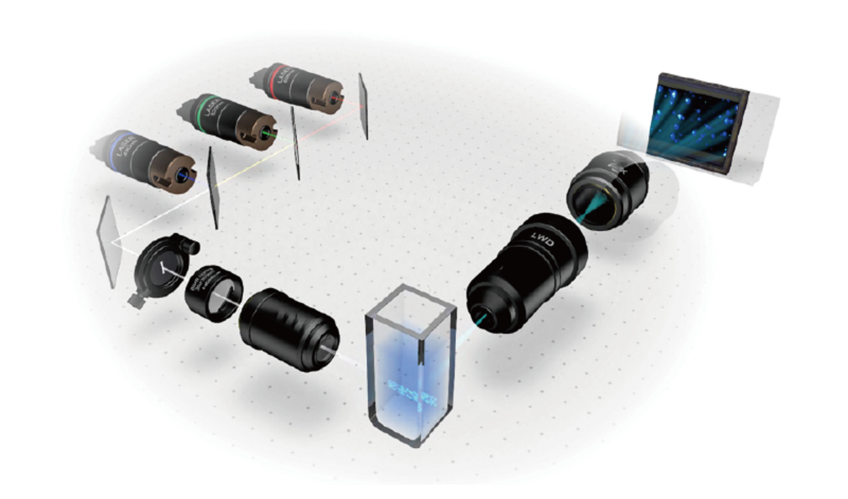
采用多波长激光对外泌体、病毒等纳米材料样品中的所有颗粒进行完整、详细的分析。
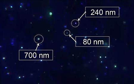
外泌体、病毒和纳米颗粒都具有较宽的粒径分布,这使得传统的纳米颗粒追踪分析 (NTA)仪无法准确测量它们的粒径分布。ViewSizer 3000的三个激光器可同时工作,可在同一样品中收集各种尺寸的*准确的分布和浓度信息。如果某一颗粒来自某一激光的散射光信号太强使检测器达到饱和,软件会自动使用来自较低功率激光器的数据来确保获得*准确的尺寸和浓度信息。另一方面,当来自某一激光的散射光信号太弱而无法检测时,软件会使用更高功率激光的数据来准确跟踪颗粒。
交叉污染是所有分析中都存在的问题。简化清洁意味着**清洁。易于拆卸的样品池可以拆卸以进行快速、**的清洁,从而获得更好的数据。
摆脱传统 NTA 的限制
准确灵敏的分析,无交叉污染
请联系我们获取更多信息
ViewSizer 通过多激光纳米颗粒追踪分析技术 (NTA) 得到颗粒粒径及粒径分布。多个激光器可分析同一样品中各种不同尺寸的颗粒,分辨率更高。
NTA 可用于对测量体积中的颗粒进行计数。该测量方法可校正粒径对有效测量体积的影响。
样品池可**拆卸,拆卸后清洗更方便**。拆卸、清洁和重新组装比冲洗流通池更快。此外,配备多个样品池可样品测量通量,也可分配给共享(核心)设施中的各个小组。
| Biological characterization using protein crystal measurements | https://bioprocessintl.com/analytical/product-characterization/biological-characterization-using-protein-crystal-measurements/ |
| A lipase-independent pathway of lipid release and immune modulation by adipocytes | https://science.sciencemag.org/content/363/6430/989 |
| Application of a novel new multispectral nanoparticle tracking technique | https://iopscience.iop.org/article/10.1088/1361-6501/aab940/meta |
| Biophysical characterization of polydisperse liposomal adjuvant formulations | https://doi.org/10.1016/j.bbrc.2020.05.156 |
| Characterisation of particles in solution – a perspective on light scattering and comparative technologies | https://doi.org/10.1080/14686996.2018.1517587 |
| Cyclodextrin Reduces Intravenous Toxicity of a Model Compound | https://doi.org/10.1016/j.xphs.2019.01.004 |
| Development and anti-Candida evaluation of the vaginal delivery system of amphotericin B nanosuspension-loaded thermogel | https://doi.org/10.1080/1061186X.2018.1434660 |
| Electrochemical sensor based on F,N-doped carbon dots decorated laccase for detection of catechol | https://doi.org/10.1016/j.jelechem.2019.03.071 |
| Light scattering by pure water and seawater: the depolarization ratio and its variation with salinity | https://doi.org/10.1364/AO.58.000991 |
| Lipid Nanoparticle-Delivered Chemically Modified mRNA Restores Chloride Secretion in Cystic Fibrosis | https://doi.org/10.1016/j.ymthe.2018.05.014 |
| Mesenchymal Stromal Cell Bioreactor for Ex Vivo Reprogramming of Human Immune Cells | https://doi.org/10.1038/s41598-020-67039-w |
| Multifunctional Nanocomposites Based on Liposomes and Layered Double Hydroxides Conjugated with Glycylsarcosine for Efficient Topical Drug Delivery to the Posterior Segment of the Eye | https://doi.org/10.1021/acs.molpharmaceut.8b01136 |
| Particle size analysis of polydisperse liposome formulations with a novel multispectral advanced nanoparticle tracking technology | https://doi.org/10.1016/j.ijpharm.2019.06.013 |
| Review of nanoparticles in ultrapure water: definitions and current metrologies for detection and control | https://www.ultrapuremicro.com/articles/review-of-nanoparticles-in-ultrapure-water-definitions-and-current-metrologies-for-detection-and-control |
| Spark erosion as a high-throughput method for producing bimodal nanostructured 316L stainless steel powder | https://doi.org/10.1016/j.powtec.2018.01.012 |
| Synthesis and Characterization of EGFR-Targeted Immunoporphysomes | http://hdl.handle.net/1807/89548 |
| Synthesis of Ultrasmall Synthetic Melanin Nanoparticles by UV Irradiation in Acidic and Neutral Conditions | https://pubs.acs.org/doi/abs/10.1021/acsabm.9b00747 |
| Nanoparticle Tracking Analysis for the Quantification and Size Determination of Extracellular Vesicles | Protocol (jove.com) | https://doi.org/10.3791/62447 |
| Isolation and characterization of EV in Saliva of Children with Asthma | https://evcna.com/article/view/3962 |
| Spinal cord injury alters microRNA and CD81+ exosome levels in plasma extracellular nanoparticles with neuroinflammatory potential | https://doi.org/10.1016/j.bbi.2020.12.007 |
| Skeletal muscle tissue secretes more extracellular vesicles than white adipose tissue and myofibers are a major source ex vivo but not in vivo | https://doi.org/10.1101/2020.09.27.313932 |
| Human milk extracellular vesicle miRNA expression and associations with maternal characteristics in a population-based cohort from the Faroe Islands | https://www.nature.com/articles/s41598-021-84809-2 |
| Purification of Cas9 - RNA Complexes by Ultrafiltration | https://doi.org/10.1002/btpr.3104 |
测量范围 | 10 nm - 15 μm |
典型样品体积 | 350 µL - 1.25 mL |
典型样品浓度(取决于样品) | 5 x 10⁶ to 2x10⁸ 颗/mL |
样品温度范围(可控) | 10° C - 50° C +/- 0.1° C |
外观尺寸 | 55 cm W x 66 cm D x 35 cm H |
重量 | 27 kg |
工作环境 | 15° C - 30° C,湿度 < 85% RH |


微信客服

服务热线

